What is the incidence of primary brain tumors in the United States?
![]()

What challenges have been encountered with conventional surgical approaches?
- Morbidity – Complications – Edema2,3,4,6,7,8
- Hemostasis Management in MIS Approaches10
- Sub-Optimal Resection Rates with Intraventricular Tumors using Ventriculoscopes15,16,17
- For Goal of Biopsy: Under-grading Disease – Sampling Error – Scant Tissue Removal9,10,11,13,14
How does the MIPS approach with NICO’s integrated systems solution overcome these challenges?
Challenge #1 – Morbidity – Complications – Edema
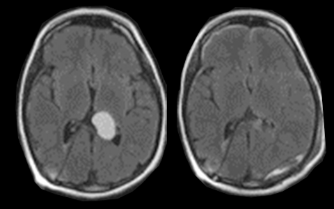
Solution: Minimally disruptive, navigable access even to eloquent areas


Challenge #2 – Hemostasis Management in MIS Approaches
Challenge #3 – Sub-Optimal Resection Rates with Intraventricular Tumors using Ventriculoscopes
Challenge #3 – Sub-Optimal Resection Rates with Intraventricular Tumors using Ventriculoscopes
Solutions: Room for Bimanual Microsurgical Technique and Automated Resection
- Both the 13.5mm and 11mm BrainPath diameters can accommodate two hands in the surgical field to enable basic hemostatic principles of microsurgery to be applied.
- Surgeons use a modified bipolar for arterial bleeding and patties with hemostatic agents and warm saline irrigation for venous bleeding.
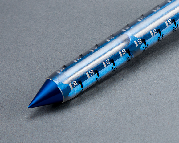
- Myriad NOVUS enables automated resection within the BrainPath corridor, especially useful on larger and/or fibrotic tumors.
![]()
Challenge #4 – For Goal of Biopsy: Under-grading Disease – Sampling Error – Scant Tissue Removal
Solution:
An effective way to preserve vasculature and fascicular anatomy while achieving a high yield of tissue collection
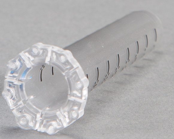
- Myriad NOVUS collects all tissue that is resected through the device for post-procedural use. Tissue is collected in a sterile, closed tissue trap that mitigates tissue degradation by limiting the sample’s exposure to the atmosphere.
- The Myriad NOVUS tissue trap holds ~ 10cc of tissue, and the clear-housing provides for easy viewing of the volume collected during the procedure.
- Myriad NOVUS resects tissue in a mechanical, non-ablative/non-thermal format, so the architectural viability of the specimen is still maintained. With the cutter engaged, the Myriad NOVUS is capable of 1,400 bites of tissue (biopsies) per minute13.
- Myriad NOVUS with a 3rd party navigation system enables correlation of resected tissue samples to intratumoral location.
- Myriad NOVUS with the tissue collection/preservation components automate and standardize the intraoperative bio-specimen collection and preservation process, eliminating the need for a full-time employee to do so.
- Myriad NOVUS with the tissue collection/preservation components allow for efficient automated collection of a greater volume of tissue while creating a biologically friendly environment for use in patient specific tissue studies and protocols.
![]()
Key Publications
Citations
- Ostrom QT et al. CBTRUS Statistical Report: Primary Brain and Central Nervous System Tumors Diagnosed in the United States in 2011–2015. Neuro Oncol (2018) 20 (suppl 4). October 2018. https://academic.oup.com/neuro-oncology/issue/20/suppl_4
- Cao, L., C. Li, Y. Zhang, and S. Gui. Surgical resection of unilateral thalamic tumors in adults: approaches and outcomes. BMC Neurol 2015; 15:229 https://doi.org/10.1186/s12883-015-0487-x
- Kelly, P. J. Stereotactic biopsy and resection of thalamic astrocytomas. Neurosurgery 1989; 25:185-94; discussion 194-5 https://doi.org/10.1227/00006123-198908000-00006
- McGirt MJ et al. Association of surgically acquired motor and language deficits on overall survival after resection of glioblastoma multiforme. Neurosurgery. Vol 65, Issue 3, 2009, 463-470 https://doi.org/10.1227/01.NEU.0000349763.42238.E9
- Chang SM et al. Patterns of Care for Adults With Newly Diagnosed Malignant Glioma. JAMA. 2005;293(5):557-564. doi:10.1001/jama.293.5.557
- Hawasli AR et al. Stereotactic laser ablation of high-grade gliomas. Journal of Neurosurgery. Neurosurg Focus. 2014. doi:10.3171/2014.9.FOCUS14471
- Pruitt R et al. Complication avoidance in laser interstitial thermal therapy. Lessons Learned. Journal of Neurosurgery, 2016, doi:10.3171/2016.3.JNS152147
- Norred SE et al. Magnetic-resonance-guided laser induced thermal therapy for glioblastoma multiforme: A review. Biomed Research International. 2014: 761312. http://dx.doi.org/10.1155/2014/761312
- Jackson RJ. et al. Limitations of stereotactic biopsy in the initial management of gliomas. Neuro-Oncology. 2001 Jul; 3(3): 193–200. doi: [10.1093/neuonc/3.3.193]
- Jackson C. et al. Minimally Invasive Biopsies of Deep Seated Brain Lesions Using tubular retractors under exoscopic visualization. Journal of Neurological Surgery. https://doi.org/10.1055/s-0037-1602698
- Khatab S et al. Frameless image-guided stereotactic brain biopsies: emphasis on diagnostic yield. Acta Neurochir. 2014. 156:1441-1450 doi:10.1007/s00701-014-2145-2
- Malone et al. Complications Following Stereotactic Needle Biopsy of Intracranial Tumors. World Neurosurgery. Citation: World Neurosurg. (2015) 84, 4:1084-1089. http://dx.doi.org/10.1016/j.wneu.2015.05.025
- Data on file. Provided by Brian Dougherty, Rose Hulman Ventures.
- Gutt-Will M et al. Frequent Diagnostic Under-Grading in Isocitrate Dehydrogenase Wild-Type Gliomas due to Small Pathological Tissue Samples. Neurosurgery, nyy433, https://doi.org/10.1093/neuros/nyy433
- Sheikh et al. Endoscopic verses microsurgical resection of colloid cysts; a systemic review and meta-analysis of 1278 patients. World Neurosurgery. 2014; 82:1187-1197
- Mohanty A et al. Initial experience with endoscopic side cutting aspiration system in pure neuroendoscopic excision of larger intraventricular tumors. World Neurosurgery. 2013; 80(5) https://doi.org/10.1016/j.wneu.2012.11.070
- Bailes J et al. Minimally Invasive Transsulcal Resection of Intraventricular and Periventricular Lesions Through a Tubular Retractor System: Multicentric Experience and Results. World Neurosurgery. 2016. https://dx.doi.org/10.1016/j.wneu.2015.12.100
- Kassam AB et al. Part II: an evaluation of an integrated systems approach using diffusion-weighted, image-guided, Exoscopicassisted, transulcal radial corridors. Innovative Neurosurgery. 2015; 3(1-2): 25-33. http://dx.doi.org/10.1515/ins-2014-0012
- Chaichana et al. Minimally invasive resection of deep-seated high-grade gliomas using tubular retractors and exoscopic visualization. Journal of Neurosurgery. J Neurol Surg A Cent Eur Neurosurg DOI: 10.1055/s-0038-1641738
- Gassie K et al. Minimally invasive tubular retractor-assisted biopsy and resection of subcortical intra-axial gliomas and other neoplasms. Journal of Neurosurgical Sciences. 2018. doi: 10.23736/S0390-5616.18.04466-1
- Day J.D., Transsulcal Parafascicular Surgery Using BrainPath for Subcortical Lesions. Clinical Neurosurgery. 2017; 64(1):151–156. doi:10.1093/neuros/nyx324