What is the incidence of cerebral cavernous malformations in the United States?
1 in 500 people have a CCM1
Majority will have no symptoms2
![]()
CCMs are the most common vascular abnormality,
making up 10 – 25% of all vascular malformations3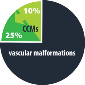
What challenges have been encountered with conventional surgical approaches?
- Safe access to deeper CM’s particularly in the insula, basal ganglia & thalamus4
- Deficits – Morbidity4,5
How does the MIPS approach with NICO’s integrated systems solution overcome these challenges?
Challenge #1 – Safe access to deeper CM’s
Solution: The MIPS approach using NICO’s systems solution provides non-disruptive subcortical access and may enable complete removal of the offending lesion as highlighted in the following publications:
TABLE 1 - Summary of Patients Treated With Minimally Invasive Parafascicular Approach Utilizing the BrainPath System (NICO Corp) for Resection of Cerebral Cavernous Malformations
| Patient | Age, sex | Presentation | Location | Complete resection | Complications | Follow-up (months) |
|---|---|---|---|---|---|---|
| 1 | 21, female | Headaches | Right frontal | Yes | None | 41 |
| 2 | 36, male | Seizure | Left frontal | Yes | Wound Dehiscence | 17 |
| 3 | 64, female | Incidental | Left Parietal | Yes | None | 10 |
| 4 | 26, female | Seizure | Right Parietal | Yes | None | 22 |
| 5 | 19, male | Seizure | Left frontal | Yes | None | 5 |
| 6 | 23, male | Seizure | Right Temporal | Yes | None | 24 |
TABLE 2 - Summary of cerebral cavernous malformation patients treated with endoport-assisted microsurgical resection
| Patient | Age (years), sex | Clinical presentation | mRS at presentation | CCM maximum diameter cm/volume (cm3) | Location | Complete resection | Complications | Follow-up (months) | mRS score at follow-up |
|---|---|---|---|---|---|---|---|---|---|
| 1 | 28, female | Syncope, tongue and arm numbness | 2 | 4.5/41.0 | Corpus callosum | No | None | 2 | 0 |
| 2 | 26, female | Headache | 2 | 0.7/0.17 | Left frontal | Yes | None | 6 | 2 |
| 3 | 47, female | Headache | 2 | 1.0/0.63 | Corpus callosum | Yes | None | 12 | 2 |
Challenge #2 – Deficits – Morbidity
Solution: The MIPS approach using NICO’s systems solution may enable preservation of neurological function as highlighted in the following publications:
- “Obviated the need for dissection of sylvian & hemispheric fissures”5
- “Tubular retractor is aided by maintaining full brain and increases accuracy of navigation…Normal to slightly elevated ICP drives lesion, particularly hematoma into sheath, limits dissection off of critical structures”5
- “The BrainPath system allows for a trans-sulcal approach which splits the white matter fiber tracts rather than cutting through them and thereby minimizes damage to the surrounding brain tissue. With preoperative DTI, the port also allows for trajectory planning avoiding white matter tracts that surround the lesion. This is particularly relevant for CM’s located in eloquent brain. When choosing the surgical trajectory, utilizing the information generated by the navigation sequence and DTI, we do not necessarily consider the shortest path from the surface to the lesion as correct one. By harnessing both technologies, the BrainPath system & DTI, we aim for the safest path that would keep both white matter tracts and gray matter intact, respectively”6
- “While endoport splitting of white matter fibers is not entirely atraumatic due to mechanical stretching tissue, the device circular nature allows more even distribution force compared to traditional retractor blades”7


Key Publications
Citations
- Mouchtouris, et al. Management of Cerebral Cavernous Malformations: From Diagnosis to Treatment. Scientific World Journal Volume 2015, ID 808314, 8 pages
- Angioma Alliance Website
- AANS website
- Awad, et al. Cavernous angiomas: deconstructing a neurosurgical disease. J Neurosurgery Volume 131. July 2019
- Amenta, et al. Resection of left posterolateral thalamic cavernoma with NICO BrainPath Sheath: Case Report, technical note and review of the literature. Interdisciplinary Neurosurgery: Advance Techniques 5 (2016) 12-17
- Goren, et al. Minimally Invasive Parafascicular Surgery for Resection of Cerebral Cavernous Malformations utilizing Image-Guided BrainPath System. Operative Neurosurgery 2018
- Ding, et al. Endoport-assisted microsurgical resection of cerebral cavernous malformations. J. Cl in Neuroscience (2015)