What is the incidence of brain METs in the United States?
Of All Cancer Patients
20 – 40%
Will Develop a Brain MET1
Annual MET Incidence2
20% Will Become Symptomatic
200,000 Cases Annually
Brain METs are the most common intracranial neoplasm in adults3
What challenges have been encountered with conventional surgical approaches?
Morbidity – Complications – Edema3-8
Increased risk of leptomeningeal spread due to ultrasonic aspirators.9,10
Hemostasis management in MIS approaches.11
Harvesting tissue in MIS approaches.
How does the MIPS approach with NICO’s integrated systems solution overcome these challenges?
Challenge #1 – Morbidity – Complications – Edema
Solution: Minimally disruptive, navigable access to eloquent areas.
BrainPath is designed for navigable, trans-sulcal access with an a-traumatic conical tip that gradually dilates the tissue during cannulation
BrainPath sheath acts as a protective portal for displaced neural tissue
Challenge #2 – Increased risk of leptomeningeal spread due to ultrasonic aspirators.
Solution: Using the NICO Myriad NOVUS the surgeon can choose to employ a pseudo en bloc technique to keep the external capsule intact while debulking the inner section of the MET.
Challenge #3 – Hemostasis management in MIS approaches.
Solution: Room for bimanual microsurgical technique.
Both the 13.5mm and 11mm BrainPath diameters can accommodate two hands in the surgical field to enable basic hemostasis principles of microsurgery to be applied
Challenge #4 – Harvesting tissue in MIS approaches.
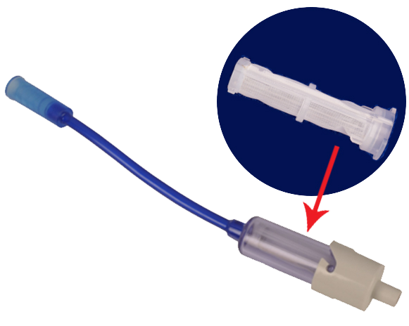
Solution: High yield tissue collection with NICO Myriad NOVUS and Automated Preservation System.
Myriad NOVUS resects tissue in a mechanical, non-ablative and non-thermal format so the cellular viability of the specimen is still maintained with minimal crushed effect.
The tissue filter of the Automated Preservation System ensures all tissue is resected through the device in a sterile, closed tissue trap that mitigates tissue degradation by limiting the sample’s exposure to the atmosphere.
Patel AJ et al. Impact of surgical methodology on the complication rate and functional outcome of patients with a single brain metastasis. Journal of Neurosurgery, May 2015. 122:1132-1143. doi:10.3171/2014.9.JNS13939
Stark AM et al. Surgical treatment for brain metastases: prognostic factors and survival in 177 patients. Neurosurg Rev. 2005, 28: 115-119. doi:10.1007/s10143-004-0364-3
Baker CM et al. Simultaneous resection of multiple metastatic brain tumors with multiple keyhole craniotomies. World Neurosrugery. October 2017. 106: 359-367. doi:10.1016/j.wneu.2017.06.118
Dubey A et al. Complications of posterior cranial fossa surgery–an institutional experience of 500 patients. Surg Neurol. 2009;72:369-375. doi:10.1016/j.surneu.2009.04.001
Hadanny A et al. Craniectomy Versus Craniotomy for Posterior Fossa Metastases: Complication Profile. World Neurosurg. 2016;89:193-198. doi:10.1016/j.wneu.2016.01.076
Brell M et al. Factors influencing surgical complications of intraaxial brain tumours. Acta Neurochir (Wien). 2000;142:739-750
Suki D, et al. Comparative risk of leptomeningeal disease after resection or stereotactic radiosurgery for solid tumor metastasis to the posterior fossa. Journal of Neurosurgery, February 2008 / Vol. 108 / No. 2 doi:10.3171/JNS/2008/108/2/0248
Ahn JH, et al. Risk for leptomeningeal seeding after resection for brain metastases: Implication of tumor location with mode of resection. Journal of Neurosurgery, May 2012 / Vol. 116 / No. 5 doi:10.3171/2012.1.JNS111560
Jackson C. et al. Minimally Invasive Biopsies of Deep Seated Brain Lesions Using tubular retractors under exoscopic visualization. Journal of Neurological Surgery. doi:10.1055/s-0037-1602698