What is the incidence of brain biopsy in the United States?
![]()
What challenges have been encountered with conventional surgical approaches?
- Invasive – Limited by lesion location due to risk for morbidity2
- Under-grading disease4 – Sampling error2,3 – Scant tissue retrieval2
- Blinded approach – Relies on the accuracy of neuro-navigation2
- Hemostasis management with no direct visualization2
- Cannot offer symptom relief – Incapable of palliative mass effect reduction
How does the MIPS approach with NICO’s integrated systems solution overcome these challenges?
Challenge #1 – Invasive
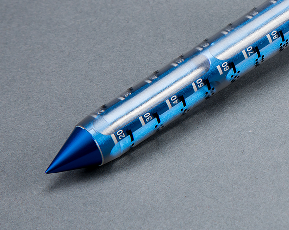
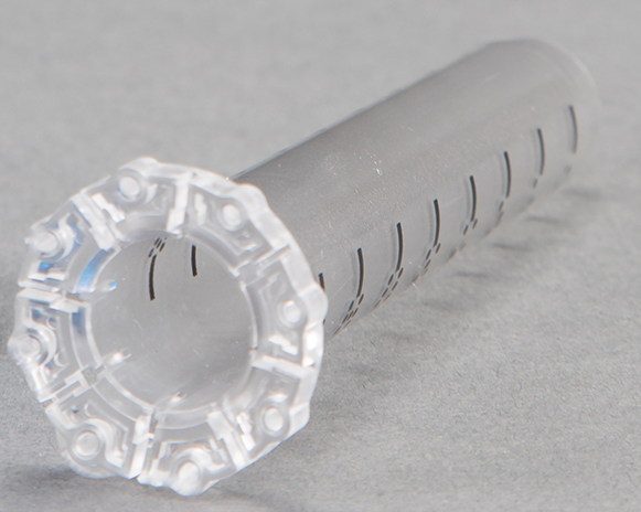
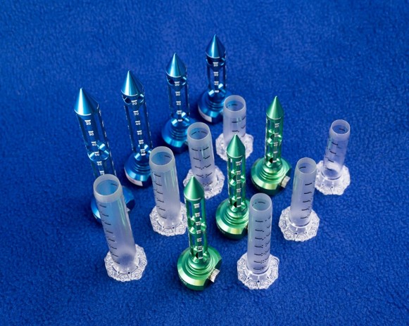
Solution: Minimally disruptive, navigable access to eloquent areas with BrainPath


Challenge #2 – Under-grading disease, sampling error, scant tissue retrieval
Solution: High yield tissue collection with NICO Myriad NOVUS and Automated Preservation System
- Myriad NOVUS tissue filter ensures all tissue resected is collected for post procedural use.
- Myriad NOVUS tissue trap holds ~ 10cc of tissue and the clear-housing provides for easy viewing of the volume collected during the procedure.
- Myriad NOVUS resects tissue in a mechanical, non-ablative and non-thermal format so the cellular viability of the specimen is still maintained with minimal crushed artifact. With the cutter engaged, the Myriad is capable of 1,400 bites of tissue per minute.
- Myriad NOVUS collects all tissue that is resected through the device in a sterile, closed tissue trap that mitigates tissue degradation by limiting the sample’s exposure to the atmosphere.
- Myriad NOVUS with the Automated Preservation System (APS) automates and standardizes the intraoperative process of tissue resection, collection and biological preservation, eliminating the need for a full time employee to do so.
- Myriad NOVUS with the APS allows for efficient automated collection of a greater volume of tissue while creating a biologically friendly environment for use in patient specific tissue studies and protocols.
Challenge #3 – Blinded approach
Solution: Direct visualization and real-time intraoperative imaging solutions
- HD optics with exoscopes have shown equal light delivery and magnification down the BrainPath sheath corridor without occupying space within the sheath.
![]()
![]()
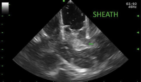
- BrainPath compatible ultrasound probes fit into the sheath corridor to make direct contact with the surgical site, which can aid in visualizing nearby anatomy surrounding and below the sheath, as well as monitor resection.

Challenge #4 – Hemostasis Management
Solution: Bimanual microsurgical technique
- Both BrainPath diameters can accommodate two hands in the surgical field to enable basic hemostatic principles of microsurgery to be applied.
- Surgeons use a modified bipolar for arterial bleeding and tamponade with hemostatic agents and warm saline irrigation for venous bleeding.5

Challenge #5 – Cannot Offer Symptom Relief
Solution: Enables Palliative Mass Effect Reduction
- The combination of BrainPath for creating access and Myriad NOVUS for automating resection enables the possibility for more of the lesion to be removed for palliative purposes, patient dependent, as compared to a needle biopsy technique.
![]()
Key Publications
Citations
- Barker FG et al. Surgery for primary supratentorial brain tumors in the United States, 1988 to 2000: The effect of provider caseload and centralization of care. Neuro-Oncology, Volume 7, Issue 1, 1 January 2005, Pages 49–63 doi:[10.1215/S1152851704000146]
- Jackson C. et al. Minimally Invasive Biopsies of Deep Seated Brain Lesions Using tubular retractors under exoscopic visualization. Journal of Neurological Surgery. https://doi.org/10.1055/s-0037-1602698
- Khatab S et al. Frameless image-guided stereotactic brain biopsies: emphasis on diagnostic yield. Acta Neurochir. 2014. 156:1441-1450 doi:10.1007/s00701-014-2145-2
- Malone et al. Complications Following Stereotactic Needle Biopsy of Intracranial Tumors. World Neurosurgery. Citation: World Neurosurg. (2015) 84, 4:1084-1089. http://dx.doi.org/10.1016/j.wneu.2015.05.025
- McLaughlin N. et al. Endoneurosurgical resection of intraventricular and intraparenchymal lesions using the port technique. World Neurosurgery. 2013; 79(2):s18: e1-38. DOI: http://dx.doi.org/10.1016/j.wneu.2012.02.022