For Immediate Release
Contact: Ryan Aspy
ryan.aspy@niconeuro.com
317.660.7118
Brain and spinal tumors glow with advanced light technology
NICO Corporation provides veterinary neurosurgeons visual contrasting capabilities with fluorescence-guided resection
INDIANAPOLIS (December 1, 2021) – Fluorescence-guided surgery (FGS) allows veterinary neurosurgeons to more thoroughly remove tumors while sparing surrounding tissues from damage. Neurosurgical medical device maker, NICO Corporation, adds new surgical adaptation with fluorescence for the NICO Myriad NOVUS®.
In this approach, patients receive fluorescein intravenously prior to surgery. During the procedure, the surgeon can toggle between the delivery of white light and the Fluorescein enabling light. Both are delivered directly at the surgical site and incorporated into the neurosurgeon’s resection handpiece.
Under the light, the margins of tumor become more pronounced, providing a clearer picture of where healthy tissue meets tumor tissue for greater accuracy, better post-surgical results for patients, and less strain for surgeons.
Prior to this breakthrough, surgeons were left to manually assess healthy tissue versus tumor. The difficulty arose in that these tissues appear as similar shades under white light, leading to eye strain, longer times in the operating room, and the possibility of leaving tumor fragments behind. Even if fluorescein was attempted in use, the light was delivered from a significant distance, often causing a washout of the filtered frequency light source.
This technology has been used by neurosurgeons and is now available for veterinary neurosurgical use. Dr. Martin Young, DVM, DACVIM (Neurology/Neurosurgery) from Bush Veterinary Neurology Service in Richmond, Va. explains, “In veterinary medicine, the masses we deal with are particularly large and removed in fragments. This system allows clear visualization of the tumor through multiple layers of removal. I have also been able to visualize small fragments of tumor that may have been missed due to location of overlying normal tissue.”
Dr. Young adds, “We have used the NICO Myriad NOVUS for removal of meningioma and glial cell tumors. Using the fluore- scein light system with the NOVUS is like using the erase function in Microsoft Paint – you just wipe away the green tumor tissue, revealing the normal blue tissue underneath.”
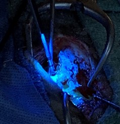
Canine brain surgery in which tumor tissue is visibly distinctive compared to healthy tissue through the use of fluorescein and a special light attached to the resection tool. Tumor is represented as the area of glowing green tissue.
The use of fluorescence-guided surgery will reshape the way neurosurgeons perform life-saving tumor removal. Access to this technology is available to veterinary neurosurgeons through Animal Health Innovations.
The NICO Myriad NOVUS pairs xenon light with the Myriad resection tool, providing precise light exactly where the surgeon needs it, improved in-situ tissue identification, non-ablative and non-thermal resection, automated tissue harvesting with the ability to annotate by intratumoral location, and biological preservation of harvested tissue for post-procedural analysis.
Learn more about the NICO Myriad NOVUS surgical system at NICOneuro.com. Follow news updates on Animal Health Innovations and NICO Corporation on LinkedIn, and stay up-to-date with NICO news and innovations on Twitter.
# # #